Saturday, 13 June 2015

SEPTIC ARTHRITIS

This is an arthritis caused by pyogenic organisms. Typically it presents as an acute painful arthirits, but it may present as subacute or chronic arthritis.
Other terms often used to describe this condition are pyogenic arthritis, infective arthritis or suppurative arthritis

Aetiopathogenesis

It is more common in children and males are more susceptible. Other predisposing facots are poor hygiene, poor resistance, diabetes etc. Staph aureus is the commonest causative organism. Other organisms are streptococcus pneumococcus and gonococcus.
The organisms reach the joint by one of the following routes:

- Haematogenous: This is the commonest route. There may be a primary focus of infection in the form of pyoderma, throat infection, septicaemia.

- Secondary to nearby oseomyelitis: This is a particularly common route in joints with intra articular metaphysis eg the hip shoulder etc

- Penetrating wounds: the knee, being a superficial joint is often affected via this route

- Iatrogenic: This may occur following intra articular steroid injections in different arthritis and during femoral artery punctures for blood collection

-Umbilical cord sepsis: in infants can travel to joints

As the organism reaches the joint by one of the above routes, there begins in inflammatory response in the synovium resukting in the exudation of fluid within the joint. Joint cartilage is destroyed by inflammatory granulation tissue and lysosomal enzymes in the joint exudate. Outcome, varies from complete healing to total destruction of the joint. The latter may result in a complete loss of joint movement (ankylosis).

Diagnosis

Mainly clinical
Usually a child
Knee is the commonest joint affected limb
In its subacute form, the parents may notice that the child is not allowing anybody to touch the joint.  He may not be moving it properly. In the lower limbs a paindul limp may be the first thing to draw attention. It may be associated with low grade fever.

On examination: 

The child is generally severely toxic with high temperature and tachycardia. The affected joint is swollen and held in position of ease. Palpation reveals increased temperature, tenderness and effusion
There is severe limitation in the joint movements in all directions. Any attempts at either passive or active movements causes severe pain and muscle spasms.  In subacute forms, some amount of movement is possible.

Investigations

Radiological Examination:
Dx in early stage is crucial.
Xrays are usually normal
A careful look at the Xray may reveal increased joint space and a soft tissue shadow corresponding to the distended capsule due to the swelling of the joint. Ultrasound examination is useful in detecting collection in deep joints such as the hip and shoulder. If found, one could aspirate the fluid and send for culturing the organism responsible for infection

In the later stagem the joint space is narrowed. There may be irregularity of the joint margins. Occasionally there may be a subluxation or dislocation of the joint.

Blood shows neutrophilic leucocytosis.
ESR in markedly elevated. A blood culture may grow the causative organisms

Joint aspiration is the quickest and the best method of diagnosing septic arthritis. The fluid may show features of acute septic inflammation. Gram staining provides a clue to the type of organism, till one gets the culture type of organism till one gets the culture report.


Differential Diagnosis

Acute osteomyelitis
Acute lymphadenitis
Acute bursitis

- mimic an arthritis because in some of these conditions, the joint is kept in a deformed position. Also there may be pain and muscle with attempted movements, but these signs are basically becasue the body is trying to prevent any because the body is trying to prevent any motion in the vicinity of the inflamed part. Careful examination reveals that reasonably pain free movements are presents  at the joint, and the movements are limited in every direction. The swelling may also be localised to one side of the joint.


Other causes of acute arthritis:

An acute septic arthritis should also be differentiated from other causes of arthritis as discussed:

Rheumatic arthritis: commonly a migratory polyarthritis, but may present with only one joint affected. The subsequent fleeting character of the arthritis, high C reactive protein levels in the serum and joint aspiration helps n its diagnosis.

Haemophilia: A past history of a bleeding disorder especially in a boy with an acute painful joint, would suggest the diagnosis. Abnormal bleeding and clotting times are helpful for confirmation.

Tubercular arthritis: It may sometimes present in a rather acute form. A past or family history of tuberculosis may be present. Joint aspiration and AFB examination may be help in its diagnosis.

Treatment

Diagnose and treat asap
Confirm or rule out diagnosis by joint aspiration
Parenteral broad spectrum antibiotics.

A combination of Ceftriaxone and cloxacillin in appropriate dises is usually given. These are subsequently changed to specific antibiotics as per aspirate cultrue and sensitivity reports. The joint must be put to rest in a splint or in traction

Whenever pus is aspirated the joint should be opened up (arthrotomy), washed and closed with a suction drain. THe same can be now done arthroscopically, As the inflammation is brought under control general condition of the patient improves, fever and local signs of inflammation subside, the joint is then gradually mobilised. Antibiotics are continued for 6 weeks.

In late cases: with radiological destruction of the joint margins movement. In such cases after an arthrotomy  and extensivedebridement of the joint. It is immobilised in the position of optimum function, so that as the disease heals, ankylosis occurs in that position.

Complications

Deformity
Stiffness
Pathological dislocation: as the joint gets filled with inflammatory exudate, the supporting ligaments and joint capsule get stretched. Muscle spasm associated with the disease may result in pathological dislocation of the joint. Posterior dislocation of the hip and triple displacement of the knee occur.

Osteoarthritis: Even if septic arthritis has been treated rather early some permanent changes in the articular cartilage occur, and give ise to early osteoarthritis a few years later.


Thursday, 11 June 2015

OSTEOMYELITIS

TYPES

Non specific organisms (pyogenic) 
 
  - strep
  - staph

Specific organisms

  -  TB
  - Syphilis
  - Salmonella
 - fungal osteomyelitis, actino and madura mycosis
 - parasitic hydatid cyst

Spread of infection

- Haematogenous

  • from a distant site in the body throat infection

- Direct

  • Atmospheric air-open fracture 

- Neighbouring focus

Mastoiditis from middle ear infection
Dental root infection producing osteomyelitis of the mandible

- Iatrogenic

Following surgery on the bone for some other reason- ORIF

Osteomyelitis is comparatively more common  in children/ infants.
If it does occur in an adult, the commonest site is the thoracolumbar spine
Other bones can be affected  in diabetes mellitus, malnutrition, drug addiction, leukaemia and other immunocompromised situations

Clinical types of OM

Acute pyogenic OM
Chronic OM
Primary subacute OM
Acute flare up of chronic OM


Pyogenic OM

  • Problem of childhood and adolescents
  • undernourished children and debilitated adults
  • common organisms- staph aureus and strep pyogenes
  • In children <4 yrs old, haemophilus influenzae
  • unusual organisms seen in drug addicts---
  • in sickle cell anaemia, they have a predilection for salmonella OM
  • Haematogenous spread is common. 
  • Focus is usually from infectiosn like impetigo, septic tooth, throat infection, infected umbilical cord
Pathogenesis

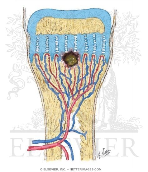
Infection commonly starts in the metaphysis of a bone
This is due to the peculiar anatomy at this zone

- arteries tend to loop causing
- vascular stasis, which
- favours colonisation

Furthermore, there is less phagocytosis in the metaphysis
                       
Organisms colonises in the bone

Causes inflammation 
Phagocytic reaction, exudation of fluid, vascular congestion

Intraosseous pressure increases
Pain, obstruction to blood flow, vascular thrombosis
Pus forms in the bone and is forced out through the Volkmann;s canal with increased intraosseous pressure to the subperiosteal space

Subperiosteal abscess


The subperiosteal abscess bursts through the periosteum into the soft tissue, forming a sinus
It may also burst into a joint causing septic arthritis

Strips the periosteum--- periosteal blood supply is lost--- sequestrum

Intramedullary extension of the pus
Intraosseous blood supply cut off 
Dead bone- Sequestrum


Small dead bone absorbed by granulation tissue and osteoclastic activity

Large gradually separated from living bone destroyed and extruded

In certain situations like hip joint the metaphysis is intracapsular and infection can easily seep into the joint

In infants and adults, vascularity pattern--- intracapsular infected epi and metaphysis---- spread to the joint

Penetrates the periosteum---- track along soft tissue and penetrates the capsule


common vascularity between epiphysis/ metaphysis and synovium

Disruption of cartilage and spread

Pathological fracture


Healing takes place at any stage
- antibiotics
- natural persistence

Healing in early stage--- exudate is reabsorbed and new bony trabeculae formed

Organism, of lesser virulence or host demonstrates increased resistance--- result in formation of persistant abscess surrounded by fibrous membrane and walled by ring of dense bone called Brodie abscess.

When sequestrum has formed exudation continues til it is absorbed or extruded

Wall off area of infection may flare up again later---- chronic OM

Clinical features

antecedent infection
irritable, restless, vomiting, high grade fever with chills
pseudoparalysis of the limb
at first, no swelling later+ infdicates subperiosteal abscess formation
affected metaphysis is tender

Fluctuation present after abscess comes to soft tissue effusion in adjacent joint- sympathetic effusion
If infection continues unabated- septicaemia- fatal termination.

Laboratory findings

- Haemoglobin: low
- Total WBC: as high as 30 000 with leucocytosis
- ESR: high
- Blood culture: demonstrates presence of bacteraemia

Radiology of APOM

- initally, up to 10 days- normal
Later:  localised areas of destruction  in the metaphysis extending ti the diaphysis- moth- eaten appearance

Periosteal elevation: multiple laminations of bone deposition parallel to the bone- appears like the onion peel appearance- seen in Ewing;s sarcoma  .

Late sign: osteoporosis with a localised segment of apparently increase density eg femoral head.

Other Investigations

MRI
CT Radiography

Diagnosis and needle aided drainage of pus for culture and sensitivity from inaccessible parts- vertebra

Radionuclide examination

Technetium phosphate- Tc99m
Positive within hours to days
initially cold spot later hot spot
depends on vascularity to the part and in the presence of increased IOP may not reach the affected part.

Gallium 67
Taken up by leucocytes
independent of vascular tree
also taken up by inflamed soft tissue and hence difficult to differentiate between cellulitis and osteomyelitis

Indium 11 labelled leucocytes
taken up by leucocytes
most suitable more sensitive
requires technical expertise and is time consuming.

MRI

Useful in early detection of osteomyelitis and soft tissue extension


DDX

Rheumatic fever: onset more gradual, consitutional symptoms less, acute and confined to the joint, polyarticular, response to salicylates and ACTH is dramatic. Antibiotics have no effect.

Ewings sarcoma: fever, leucocytosis, subperiosteal reaction- onion peel appearance, destruction confined to the diaphysis, responds to radiotherapy, biopsy shows tumour cells.

Acute septic arthritis:  fluid accumulation in the joint occurs earlier, pain and inflammation limited to the joint, joint movements grossly restricted, aspiration reveals purulent synovial fluid

Treatment: Early diagnosis with a high degree of suspicion is beneficial and necessary
Blood for investigations are collected and high doses of antibiotics are started as early as possible.

Antibiotics are changed later if necessary as per culture and sensitivity  reports

Immediate drainage is of paramount importance and is done before signs of subperiosteal infection is seen.

To wait is to invite trouble and disaster. Maximum waiting period allowed is 24 hours SOS to improve general condition patient.
Immediate drainage by opening a cortical window at the suspected site.

Closed continuous drainage for 24-48 hours.
Or leave the wound open and allow secondary healing

Prolonged antibiotics as per culture and sensitivity report for minimum period of 6 weeks with initial 2 weeks of parenteral antibiotics.

COMPLICATIONS

Acute osteomyelitis invariably ends up as chronic osteomyelitis
Septicaemia and fatal end
Multifocal osteomyelitis in debilitated individuals- rare

After an attack of acute osteomyelitis recurrence of infection is a rule- the interval may vary.
Once an osteomyelitis always an osteomyelitis

Acute OM leading to chronic OM
Haematogenous infection with a low virulence organism may be chronic from the beginning
Infection from an external wound usually causes chronic osteomyelitis

Pathology

Repair when incomplete persistance of infection
Repair process- hyperaemia formation of granulation tissue and aborption of necrotic cancellous ad cortical bone.

When sequestrum is small and infection is controlled sequestrum gets resorbed
When large and infection persists- it separates out and lies in a cavity
The surrounding tissue attempts to wall off infection--- forms thick bony wall--- called the involucrum

Pathology

Involucrum has multiple openings called cloacae, openings for exudate, debris and sequestra to drain though the sinus.
Once sequestrum  is extruded- infection is better controlled and settles down.

TYPES OF SEQUESTRUM

STIR BCD

Sandy   ------    tuberculur OM of the vertebra
Tubular ------   tubercular OM of the long bone
Ivory ---------   syphilitic osteomyelitis
Ring ----------  stump/ skeletal traction
Black ---------  prolonged exposure of bone
Cortical ------   pyogenic OM adults
Diaphyseal ---  pyogenic OM- children


Clinical features

In the period of inactivity no symptoms
Fever, pain, swelling and tenderness of bone
Sinuses discharging pus and bony spicules- sequestrum
Bone is thick, irregular or may be deformed
Skin dusky thin and scarred.

Muscles are scarred and contracted- produce deformities of adjoining joints

Radiology

Moth eaten appearance
Osteoporotic bone
Sequestrum
Involucrum
Bone thick and irregular
Bone may be deformed
Pathological fracture

Other Investigations

Sinogram

Traces the sinus tract and helps planning of surgery and removal of entire tract to prevent recurrence.

Sequestrectomy and saucerisation
Scondary healing of wound
Prolonged antibiotic- minimum of 6 weeks to 3 months according to culture and sensitivity report with initial parental antibiotics.

COMPLICATIONS
Acute flare up chronic osteomyelitis
Squamous cell carcnima of chronic discharging sinus
Contracture of muscle producing deformity of joints

Stimulation/ destruction of growth plate leading to discrepancy of limb length or deformities
Pathologic Fracture
Septic arthritis of adjacent joint- deformity and ankylosis
Amyloidosis

Brodie's abscess

Described by brodie in the tibial metaphysis in 1832
Indicates subacute pyogenic osteomyelitis usually of staphylococcal origin
Commonly seen in children- boys

Commonly affects the ends of the tibial bone
ABscess varies from 1cm-4cm in diameter
Radiologically seen as cavity surrounded by dense ring of bone.

Appropriate antibiotics may decrease the size of the lesion
If pain persists- may require surgical decompression of the abscess.


Wednesday, 10 June 2015

EXAMINATION OF HIP

Wash your hands
Introduce yourself with your full name and title, confirm that you have the correct patient with their name and date of birth. Explain what you want you want to do and obtain informed consent
Request for chaperone
Ask for pain and if need painkillers
Ensure privacy and adequately expose  the patient. If the patient is female, maintain her modesty

Check for walking aids around the bed and supports as such. Ask the patient to stand up and walk away from you and back.
Describe the gait of the patient.

From the front, with the patient standing in front of you, squat down to the level of the hips.


Observe for stance, shoulder and pelvis parallel to the floor.

What are the level of the shoulders: may indicate the presence of limb length discrepancy
Deformities
Muscle wasting

From the side

Is there any evidence of the deformity in the spine? Normal spinal curvatures and deformities in the spine like lordosis and kyphosis
Trochanteric area: examine for any swelling.

On the lateral aspect of the hip and limb: swellings, scars, sinuses, skin changes

From the back:

- Deformities in the spine : scoliosis
- Tell tale signs in the back: evidence of spinal deformities
- Levels of the posterior superior iliac spines/ iliac crests
- Sacral dimples- levels of the sacroiliac joints
- Level of the gluteal and thigh folds: indication to limb length discrepancy
- Any gluteal atrophy
-On posterior aspect of hip and limb- swelling, scars, sinuses
- Look for evidence of deformity if seen

Now since she is still standing we can straight away do the Trendelenburg instability test, or else wait til later to do it

Trendelenburg test:  to demonstrate an ineffective abductor mechanism (gluteus medius weakness, dislocated or destroyed head of femir, nonunion of neck of femur, coxa vara).

Kneel in front of the patient and hold their pelvis. Ask the patient to alternatively  stand on one leg. Normally the iliac crest on the side raised should rise. If it falls, it may be due to weakness of gluteal muscles

Ask the patient to lie down flat on the coach. From the front with the patient supine


  • Attitude of the limb: position of various joints, direction and degree

flexed, abduted internally rotated--- posterior dislocation

externally rotated--- fracture neck of femur.
  • Deformity of the hip if any and details therein
  • Spine: evidence of exaggerated lumbar lordosis: level of shoulders
  • ASIS: level of anterior superior iliac spines: abduction/ adduction deformities
  • Femoral triangle examination: swelling and its description
  • Presence of swelling scars and sinuses, dilated veins and description  of each as on inspection of the hip and entire limb. 
  • On the side. see the trochanteric area. Confirm with the knee flexed.
  • Trochanteric bursa- inflamed--- swelling/ fluid/ painful
  • Muscle wasting
  • Limb length discrepancy
    • Galleazi's sign: flex knees to 90 degrees and hips and ankles at 45 degrees. Put malleoli at same level. Note level of knees. Parallel level of femurs and tibia. 
    • If the knees are at different levels, but tibia is parallel so discrepancy not above knee (ie its in tibia). 
    • If femurs are parallel, discrepancy not below knee ie in femur. 
Palpation

Local rise of temperature over the area of interest. Check the normal side
Tenderness--- at the joint line--- by the side of the femoral pulse.
If it is very tender, do it gently. Do not make the patient cry

If cannot feel femoral pulsation- is it dislocation?
= flexion deformity
= internally rotated
= shortenimg

Tenderness over the greater trochanter- trochanteric bursitis

Confirm all inspectory findings in the spine and back- regarding spinal deformities, swelling, scars, sinuses, limb length discrepancy
Look for presence of lymph nodes whenever appropriate
Bony prominences: ASIS, greater trochanter (height, tenderness, width, surface of he greater trochanter in comparison to the opposite side), patellar height and medial malleolar level. 
Bony irregularities: look for presence of bony irregularities and details of where such irregularities are palpated. Also look for any deformity of the bone and record direction of the deformity
Palpate for all peripheral pulses and look for motor and sensory disturbances in areas concerned
Vascular sign of Narath: in condition where the head or neck of the femur are not in place of are destroyed as a complication of a disease process, the femoral pulsation on the affected side is well felt. 

When increased lumbar lordosis is present: indicates primary spinal deformity or an underlying fixed flexion deformity of the hip.
The degree of deformity has to be measured in degrees by doing the Thomas test. When ASIS is found to be at different levels, it indicates spinal deformity leading  to pelvic obliquity or a fixed abduction or adduction deformity of the hip leading to obliquity of the pelvis. When ASIS on the affected side is at a lower level it indicates an abduction deformity on the hip and when at a higher level indicates an adduction deformity of the hip.

The degree of deformity has to be  measured only after squaring the pelvis and unmasking the deformity. Deformity has to be quantified in degrees. 

Movement
First do Active Movement, then only do passive movement. 

You may put your left hand  under the patient's spine to check  if the patient is using the pelvis  or spine during the movement phase. 
Ask the patient to first do flexion which is normally up to 120 degrees. See if any pain.

Ask the patient to turn prone to check extension. Make sure you place the hand on the pelvis to fix it. Normal range is from 0-20 degrees. 


Square the pelvis by placing your left hand over the patient's left hip.

Adduction: bring the foot to the opposite side, fully extended.  25 degrees from midline


Abduction: with the patient's leg fully extended, bring the leg closest to you 0-45 degrees from midline
Repeat for the opposite leg.


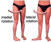
Internal rotation and external rotation. Repeat for other leg. Normal range is 25-45 degrees in and out.
Image result for hip internal rotation

Flex the knee and hip  to 90 degrees. Move the foot inwards for internal rotation and outwards for external rotation. 

All the ranges of movements in all directions normally possible in the joint has to be measured in degrees.

Movements have to be recorded in a tabular column showing the movements on the right hand and the left sides in each direction and a column to record the difference foind in each directions. 

This tabular column  can also depict  the power in each group of muscle eg flexors, extensors, abductors etc.
When fixed deformities are found
Movement in opposite direction is not possible.
Movements in the same direction as the deformity may be possible. 

Eg when  fixed abduction deformity is seen to be present, adduction at the hip will not be present and vice versa. 

ALL MOVEMENT SHOULD BE RECORDED AS RANGE OF MOVEMENT WITH A DEFINITE START AND END POINT MENTIONED IN DEGREES.


Measurements

All bony prominences should be marked with a marking pen on both  side before measurements
The tape should be placed gently on these points during measurements. Care should be taken to ensure that the points have not been moved under the examining fingers.


Apparent length: from the xiphisternum to the medial malleolus with the patient supine and lying down as straight as possible. In abduction  or flexion deformity  there may be apparent shortening.

True measurement of the entire limb in genuine shortening of the limb

1) Square the pelvis
2) Both limbs to be kept in similar positions when measuring
3) Measure from ASIS to the medial malleolus on each side

Segmental true measurement

1) Squared pelvis
2) Femoral length

- ASIS to the medial joint line
- The medial joint line is palpated with the knee in the 10-0 degrees of flexion when possible. The tibial tuberosity is identified and the examining fingers are un proximally on the tibia along the flare of the tibia
- A depression is felt as the fingers run proximally which is placed perpendicular to the axis of the femur on the medial and lateral aspects of the knees. This represents the medial and lateral joint line
This is confirmed by then moving the joint into flexion and extension whenever possible with the examining fingers still placed on the medial and lateral aspects of the knee. The movements are appreciated  to be occurring at that level confirming it to be the joint line. 

Tibial length
From medial joint line to the medial malleolus

Limb girth

If there is evidence of muscle wasting. 

Bryant's triangle: to decide if there is supratrochanteric or infratrochanteric shorting.1st draw  a line joining the ASIS and the greater trochanter. Then one line is dropped perpendicularly to the bed from the ASIS. Join a line to that line. Compare with opposite side. If it is shorter, it signifies supratrochanteric shortening.


Shoemaker's line: 


 Nelaton's line: 


Special Test

Thomas test
DO NOT DO ON ANYONE WHO HAS HAD HIP REPLACEMENT
With the patient supine on a hard surface, look for evidence of excessive lumbar lordosis by looking for light passing through and through the lumbar area between the back and the couch

Pass the palm of the hand under the lumbar region. This cannot be done in a normal situation. If the palm can be passed  under the lumbar area between the back and the couch it indicates excessive lumbar lordosis

With the palm under the lumbar back, flex the hip on the unaffected side through its full range of flexion and continue to flex it beyond till the lumbar back just touches the hand. Notice the hip on the affected side flexing with this manoeuvre. 

With the unaffected hip in position of flexion, passively extend the hip on the affected side as much as possible without allowing the patient to arch the back. The angle that the back of the thigh makes to the couch on the affected side is the amount of fixed flexion deformity of the hip. 
Incomplete extension quantify fixed flexion deformity in the hip.


SUMMARISE

Inspect for 
walking aids
attitude
SSSW
deformity
shoulder tilt or lumbar lordosis
trendelenburg test
gait


Palpate for

temperature
tenderness
 ASIS
 Iliac crest
 PSIS
 Sacroiliac joint
 greater trochanter
 Greater trochanter
 Adductor area
  
Bryant's triangle
Shoemaker
Nelaton


Measurement

Apparent length
Square pelvis for true length
Muscle wasting

Movement

Flexion 0-140
Extension 0-20
Internal rotation  0-30
External rotation 0-40
Abduction  0-45
Adduction  020
Extension 0-15

Special test:

Thomas test


Random Notes

Hip is innervated by L2-S1
L2 also innervates the knee, hence hip pathology may have referred pain in the knee
Spine gives off L3

How to locate the hip- femoral triangle
Squaring of pelvis--- why is it called squaring?

iliac crest is on the same level.Limbs in same position. 

Abduction- shortens the limb
Adduction- lengthens the limb.

What are causes of true shortening of limb?

= fractures
= hip arthroplasty
= slipped upper femoral epiphysis
= hip dislocation
=  juvenile osteoarthritis
= septic arthritis
= loss or articular cartilges
= congenital coxa vara
= growth disturbances.


Patient cannot abduct and internally rotate. What do you suppose it is?

Osteochondritis of femoral head- Perthes disease.

Tuesday, 9 June 2015

Avascular Necrosis of Bone

This is bone loss due to loss of vascularity of the bone after a fracture or dislocation. May be spontaneous also.
It is a type of osteonecrosis.
Bone death or severe marrow and bone cell damage due to decrease in blood supply.

Sites:

- Femoral head
- Scaphoid
- Talus
- Lunate
- Head of humerus

Pathogenesis

Arterial insufficiency
- Fracture
- Dislocation

Venous occlusion
- Perthes disease

Intraosseous capillary compression

  • Infection
  • Gaucher;s disease - cortisone, alcohol
  • Caisson's disease

Intravascular capillary occlusion

Articular space normal
  • Sickle cell disease
  • Alcohol
  • Caisson disease
  • Corticosteroid
Pathology

Changes begin within a week of the insult and evolve over a period of 2-4 years. 
Pathology same in all conditions whatever the cause + features of underlying disorder
Bone cells die in 12-48hrs

Necrotic segment gets demarcated and granulation tissue grows in

New bone is laid down on the dead: increase in mineral mass: seen as sclerosis on Xray

With weight bearing: structural failure in new bone

Cracking of subchondral bone

Fragmentaation of necrotic bone/new bone

Destruction of joint surface

Is avascular necrosis reversible? Does complete healing occur? yes

Clinical Features

  • Pain: in or near a joint.
  • Only in certain movements
  • Tenderness/ swelling
  • Only in the superficial bones
  • Clicks in the join
  • Loose articular surfave
  • Stiffness of joint
  •  In later stages of the disease
  • Movements: one plane restricted early on
  • Advanced cases: fixed deformities. 
Radiology
Increased bone density
Subchondral fracture
Distortion of articular surface

Staging: Ficat & Arlet

Stage I: Pre-clinical phase: no radiological abnormality
Stage II: Early sclerois, bone contour normal
Stage III: Sclerosis, structural damage, bone contour altered
Stage IV: Collapse or articular cartilage and secondary OA

Other Investigations
Radionuclide 

- AVN seen as cold spot
- Surrounding area seen as hot spot

MRI

- identifies  in preclinical phase

Treatment

Factors influence natural course

= type of ischaemia
= area affected
= stage of disease
= patient's age
= capacity for bone repair
= persistence of etiological  agent. 

Treatment

Early stage (Stage I/II)
Bone contour normal, small lesions
Weight bearing bone vs non weight bearing

Non operative Rx

Traction: spasm decreases

Operative Rx: Eliminate etiology

Surgery: better results

Core decompression
Muscle pedicle bone grafting
Defunctioning osteotomy

Treatment
Late stage III/IV

Structural damage and articular distortion present

Operative treatment
- Pain/ loss of function: should demand for the procedure
- Realignment osteotomy
- Arthroplasty
- Arthrodesis

CRYSTAL DEPOSITION DISEASE


This is a group condition characterised by presence of  crystals in and around joints, bursae and tendons

- Gout
- Calcium pyrophosphate dihydrate
-  Hydroxyapatite


Consequence: Crystal deposition
Remains intact and asymptomatic
Induces acute inflammatory reaction
Results in slow destruction of the affected tissue


1. Asymptomatic hyperuricaemia
2. Acute gouty arthritis
3. Intercritical gout
4.Chronic tophaceous gout
5. Miscellaneous: Gouty nephropathy


Gout:
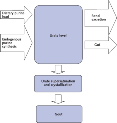
Definition: A disease characterised by hyperuricaemia and formation of monosodium urate crystal deposition in the body.
Gouty arthritis is joint inflammation caused by MSUM crystals in synovial fluid and joints.


Disorder of purine metabolism


Uric acid is completely filtered by the glomerulus
Completely (essential) reabsorbed in the proximal tubule
Approximately 50% is seceted back into the tubule in the descending loop
Approximately 80% of that 50% is reabsorbed in the ascending loop.
Net excretion=10% of filtered load


Common in males M:F=20:1
In females after menopause
Hyperuricaemia


Monosodium urate monohydrate crystals
Recurrent acute synovitis

Two types:

Primary: 95%
Inherited disorder
over-production or under of uric acid
Secondary 5%
Acquired conditions

Myeloproliferative disorders---- overproduction
Renal failure----- under excretion

Gout:

All patients with high serum uric acid do not develop gout
Crystals are deposited in minute clumps- remain inert for years
Local trauma- disperses crystals into joint- acute inflammatory reaction
Each crystal- phagocytosed or float free in joint.

Over years urate deposits build up in joints, periarticular tissue, tendon and bursa- destroy articular cartilage and periarticular bone

MTPJ of great toe
Olecranon bursa
Pinnae of ears

Tophi varies in size <1cm to several cm.
It may ulcerate through the skin

Clinical Features

Men >30 years of age
Women after menopause
Obese, alcohol friendly
Patients on diuretics which increases tubular reabsoption of uric acid
Family history


Acute gout

Sudden severe joint pain precipitated by

- Minor trauma
- Surgery
- Minor illness
- Unaccustomed exercise
- Alcohol

More in the night
Lasts for 1-2 weeks and completely resolves

Common sites are affected
Joint hot and tender DDx: septic arthritis
Hyperuricaemia not diagnostic
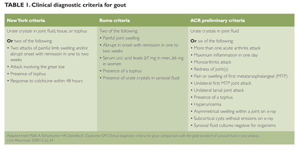
Diagnosis confirmed by synovial fluid analysis for negatively birefringent urate crystals


Chronic gout

Recurrent attacks leads to increased joint stiffness and deformity

  • Tophi appear over the common sites
  • Large tophi may ulcerate the skin- chalky material discharged
  • Renal calculi, renal parenchymal disease
Acute Gout
Only gout

Chronic gout

Tophi appear as punched out lesions (cysts) in the para-articular bone ends
Secondary osteoarthrosis
Tophaceous disease more likely to occur in patients with

  • polyarticular presentation
  • Serum urate level>540umol/L (>9mg/dL)
  • Disease onset at younger afe (<40yrs)
Sites of tophi

- Digits of hands and feet
- Pinna of ear (classic, less common)
- Bursa around elbows and knees
- Achilles tendon


DDx:

Infection
  • Cellulitis
  • Septic arthritis
  • Infected bursitis
Reiter's disease
  • History is more protracted
  • Response to NSAIDS is less
Pseudogout
  • Affects larger joints
  • More common in women
  • Pyrophosphate dihydrate crystal deposition
Rheumatoid arthritis

  • Polyarticular gout- affecting fingers
  • Elbow tophi- mistake for subcutaneous nodule
Biopsy establishes the diagnosis


Investigations

Specific investigations for confirmation
- Serum uric acid
- Joint aspiration and crystal identification

To detect medical conditions associated gout or hyperuricaemia
- FBC
- Serum creatinine/ urea
- Serum blood glucose
- Fasting lipid profile
- UFEME
- 24hr urinary urate excretion
  • Useful if renal calculus procen to be urate stone
  • indicated if on uricosuric agent
  • assess risk of stone
  • help to indicate whether overproduction or underexcretion of urate
  • range 2:4 mmol/24hr or 0.34- 0.67g/24hr
Skeletal Xrays

Acute gouty arthritis: Normal, soft tissue swelling
Chronic tophaceous gout: tophi erosive bone lesions (punched out lesions), joint space is preserved until late stage, pathognomonic in foot and big toe.


To detect complications
- Renal imaging
- Skeletal Xrays


Acute Stage

- Rest to the joint
- NSAIDS
- Colchicine less effective
- Aspiration to relieve tense joint
- Intra articular hydrocortisone

Treatment

Interval therapy
- Loose weight
- Reduce alcohol
- Eliminate diuretics
- Uricosuric drugs--- allopurinol

Interval therapy indicated

Acute attacks occur at frequent intervals.
Presence of tophi
WHen renal function is affected
Uricosuric drugs used if renal function is normal
Allopurinol- Xanthine Oxidase inhibitor 
    NEVER STARTED IN ACUTE STAGE
    ALWAYS COVERED BY NSAIDS/COLCHICINE AS THEY WILL OTHERWISE PRECIPITATE  AN ATTACK OF GOUT

Chronic gout

Allopurinol is the drug of choice
Ulcerative tophi may have to be evacuated.


Q, What is intercritical gout

Asymptomatic period between attacks


Q. What is urate/ gouty nephropathy

Acute urate nephropathy

Urate crystals---- renal tubules---- obstructive ARF
Dehydration, low urine pH are precipitating factors

Chronic urate nephropathy

Urate crystals--- interstitium and renal medulla----- inflammation+ surrounding fibrosis----- irreversible CRF
Renal impairment can occur in 40% of chronic gout

Urate nephrolithiasis

Stones----- flank pain/ ureteric colic/ hematuria
Urate (radioluscent) or mixed calcium oxalate and/or calcium phosphate (radio-opaque)
Contributing factors: hyperuricosuria, low urine output, acidic urine
Urinary alkalinisation (potassium citrate or NaHCO3)--- dissolution of existing stones and prevention of recurrence.